1.基本原理
大多數EIT系統採用的都是如圖1所示的測量方式,數字1-16所標示的為貼上在人體表明的電極,EIT硬體系統先在電極1-2處施加一激勵電流,並在3-4,4-5,……,15-16處測得其電壓,然後切換激勵電極,改在2-3處施加一激勵電流,並在其餘的電極處測得對應的電壓,激勵電極切換一圈後,在其他電極處所測到的所有數據為一幀數據,通過測量兩幀不同時刻的數據,重構出這兩個時刻的電阻抗分布的變化。
 動態電阻抗斷層成像
圖1 EIT測量示意圖
電流激勵時,如圖 1所示,從相鄰兩個電極進行電流激勵的方式稱為鄰近激勵,從相對兩個電極(如1-9電極)進行電流激勵的方法稱為對向激勵,由於不同電流激勵方式會在內部產生不同的電場分布,從而會影響EIT信號的檢測能力和圖像的重構質量。
2.設備組成
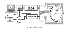
EIT系統的結構如圖2所示,由電流源、多路開關、電壓放大器、AD轉換器、單片機等共同組成。
 動態電阻抗斷層成像
圖2 EIT設備結構圖
電流源基於數字合成技術設計而成,由控制及接口模組、程控時鐘發生器、存儲單元、地址信號發生器、數模轉換器、平滑濾波器、電壓電流轉換器共同組成。其工作原理是:在微程式控制器控制下,現場可程式邏輯器件中的時鐘控制接口設定好時鐘的頻率,所產生的時鐘信號被送往計數器,計數結果作為定址信號被送往存儲單元,從而使存儲單元周期性地輸出數字量。這些數字量是對一個周期正弦信號的點等間隔採樣值,它們被依次送往DAC,再經平滑濾波後便可合成正弦波信號。此信號經電壓電流轉換器形成激勵信號。
由於EIT數據採集過程中,需依次從多個電極中選取一對作為激勵電極,並依次選取一對相鄰電極進行測量。為適應不同激勵測量模式的要求,數據採集系統應具備從這多個電極中任選兩個進行激勵,同時任選兩個進行測量的能力。可採用多路多選1模擬電子開關構成了多路開關模組。多路電子開關中的兩路的公共端分別與恆流源輸出的正負極相聯,另兩路的公共端則分別與測量通道的同相和反相輸入端相聯。
電壓測量模組需有高精度、低噪聲、寬動態範圍和寬頻帶等特性。為滿足這些要求,電壓測量模組主要由儀表放大器加程控增益放大器兩大部分構成。
由於解調模組採用數字解調方式,需要在一個信號周期內對被測信號進行多次採樣,因而所選AD轉換器應具有較高的轉換速度和較高的轉換精度。
3.圖像特點
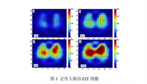
原始EIT圖像顯示的是按有限元剖分單元繪製的各單元上的阻抗相對變化值,原始EIT圖像可以經過進一步的插值平滑處理成由一定數目不同偽彩色表示的像素矩陣,如圖3所示。相對於CT、MRI圖像而言,EIT圖像解析度較低,通常為32×32、64×64、128×128像素不等。
 動態電阻抗斷層成像
圖3 正常人肺部EIT圖像
EIT圖像的不同顏色反映的是組織或器官相對阻抗變化,阻抗變化越大,圖像對應區域的顏色變化越深。雖然EIT圖像空間解析度較低,但是阻抗解析度非常高,在較好硬體系統支持下,可以對阻抗變化0.1%的目標進行成像。EIT成像的這一特點,使得它具有對微弱病變敏感,可早期發現病情的特點。
EIT圖像另一重要特點是成像速度快,通過對重構矩陣的預先計算,EIT圖像可以做到實時成像(25幀/秒以上),加之技術本身的無創無害,因此不僅可以對呼吸等快速變化進行實時成像,還能進行長時間的圖像監測。
雖然EIT圖像值目前還沒有國際統一單位,仍採用相對變化值(Arbitrary Unit),但是針對不同的臨床套用,已經提出了感興趣變化、全局變異係數等許多成像指標,有些指標已成功指導臨床的疾病診治。
4.優勢缺點
(1)優勢
EIT圖像屬於功能圖像,具有靈敏度高、安全性好、成本低廉、連續成像等優勢,可以觀察到疾病的早期變化,可以持續對功能及病情變化進行評估。
(2)缺點
EIT圖像解析度較低,干擾較大時會引入偽影。
5.醫學套用與發展前景
電阻抗斷層成像技術,特別是動態電阻抗斷層成像技術,已經展現出了良好的套用前景,主要集中在:乳腺癌檢測成像、腹部臟器功能成像、肺部呼吸功能成像、腦部功能成像等。由於臨床套用需求不同,面臨的技術難點也不同,EIT在上述領域的臨床套用研究的成熟度也不同,有的套用點已經獲得了產品認證,有的仍處於實驗室研究階段。但總的來說,近年來EIT技術越來越受到臨床研究人員的關注。
(1) 乳腺癌檢測成像
乳腺癌是臨床常見腫瘤,若發現於早期,則治癒率高,預後較好。又由於腫瘤組織具有電特性,特別是惡性腫瘤組織的電特性與周圍正常組織的電特性存在明顯區別,因此乳腺癌檢測是電阻抗成像領域關注的重點方向之一, 研究結果表明,EIT、X線鉬靶、超聲總體敏感性沒有顯著性差異,EIT可以成為X線鉬靶和超聲的有力補充, EIT結合已有技術可以提高乳腺癌早期小腫瘤的檢出率。
(2) 腹部臟器功能成像
在腹部臟器功能成像研究領域有望成功套用的研究主要為腹腔內出血監測和胃動力監測。利用電阻抗成像技術已成功監測小豬腹膜內和腹膜後出血,並由臨床套用個案報導。
(3) 肺部呼吸功能成像
呼吸是人體重要的生命體徵之一。由於空氣不導電,因此呼吸時肺部會發生較大的電特性變化,加之呼吸具有特殊的周期性時域特性,因此電阻抗成像技術在肺部病情監測中的套用研究最為廣泛,臨床成熟度也最高。肺部電阻抗成像在人工通氣、局部肺栓塞、局部肺灌注等領域都具有較好的臨床套用前景
(4) 腦部功能成像
由於腦科學研究的前沿性和腦血管疾病發生的致命危害性,因此進行顱腦電阻抗成像的相關研究具有非常重要的理論價值和套用價值。但由於顱骨具有非常大的電阻率,且在顱骨與腦實質之間又存在電阻率非常小的腦脊液層,這種空間結構上電阻率的巨大差異使得激勵電流難以穿透顱骨,腦實質內的病變反映到皮層的信號發生較大衰減,使得顱腦電阻抗成像成為這個領域最富挑戰性的難題。在腦部成像方面,已經在出血和缺血性腦卒中動物模型上獲得了穩定的成像結果,完成了硬膜下血腫鑽孔引流術監測,並進一步在腦水腫病人甘露醇脫水監測等方面取得了臨床套用。
總之,EIT成像技術在臨床的定位並不是取代CT、MRI等現有影像診斷技術,而是利用靈敏度高、安全性好、成本低廉、連續成像等優勢,彌補現有影像技術的不足,作為現有技術的有力補充,提高臨床診治能力。
擴展閱讀
[1] Panagiotis K, Montaserbellah A, Panos L.Stimulation and measurement patterns versus prior information for fast 3D EIT: A breast screening case study[J]. Signal Processing, 2013, 93(10):2838-2850.
[2] Zou Y,Guo Z.A review of electrical impedance techniques for breast cancer detection[J].Medical Engineering & Physics,2003,25(2):79-90.
[3] Sachin P N, Houserkova D, Campbell J. Breast imaging using 3D electrical impedance tomography[J]. Biomedical Papers-Olomouc, 2008, 152(1):151-154.
[4] Raneta O, Bella V, Bellova L, et al. The use of electrical impedance tomography to the differential diagnosis of pathological mammographic/sonographic findings[J]. Neoplasma, 2013, 60(6):647-654.
[5] Forsyth J, Borsic A, Halter R J,et al. Optical breast shape capture and finite-element mesh generation for electrical impedance tomography[J]. Physiological Measurement, 2011, 32(7):797-809.
[6] Shuai W J, You F S, Zhang W, et al. Image monitoring for an intraperitoneal bleeding model of pigs using electrical impedance tomography[J]. Physiological Measurement, 2008, 29(2):217-225.
[7] Shuai W J, You F S, Zhang H Y, et al. Application of electrical impedance tomography for continuous monitoring of retroperitoneal bleeding after blunt trauma[J]. Annals of Biomedical Engineering, 2009, 37(11):2373-2379.
[8] You F S, Shi X T, Shuai W J, et al. Applying electrical impedance tomography to dynamically monitor retroperitoneal bleeding in a renal trauma patient[J].Intensive Care Medicine, 2013, 39(6):1159-1160.
[9] Maximilian S S, Viktoria W, Bea B, et al. Electrical impedance tomography during major open upper abdominal surgery: a pilot-study[J]. BMC Anesthesiology, 2014, 14:51.
[10] Sarker S A, Mahalanabis D, Bardhan P K, et al. Noninvasive assessment of gastric acid secretion in man—application of electrical impedance tomography (EIT)[J]. Digestive Diseases and Sciences, 1997, 42(8):1804-1809.
[11] Pavel D, Anatolij T, Josef P, et al.Assessment of regional ventilation with the electrical impedance tomography in a patient after asphyxial cardiac arrest[J].Resuscitation, 2014, 85(8):115-117.
[12] Zhao Z, Fischer R, Frerichs I, et al.Regional ventilation in cystic fibrosis measured by electrical impedance tomography[J]. Journal of Cystic Fibrosis. 2012, 11(5):412-418.
[13] Karsten J, Bohlmann M K, Sedemund-Adib B, et al.Electrical impedance tomography may optimize ventilation in a postpartum woman with respiratory failure[J].International Journal of Obstetric Anesthesia, 2013, 22(1):67-71.
[14] Patricia B, Michael M, Helmut H, et al.Imaging of local lung ventilation under different gravitational conditions with electrical impedance tomography[J].Acta Astronautica, 2007, 60(4): 281-284.
[15] Suchomel J, Sobota V. A model of end-expiratory lung impedance dependency on total extracellular body water[C]// XIV Conference on Electrical Impedance Tomography. Heilbad Heiligenstadt, Germany: IOP Publishing, 2013:1-4. .
[16] Ferrario D, Grychtol B, Adler A,et al.Toward morphological thoracic EIT: major signal sources correspond to respective organ locations in CT[J]. IEEE Transactions on Biomedical Engineering, 2012, 59(11):3000-3008.
[17] Holder D S. Feasibility of developing a method of imaging neuronal-activity in the human brain—a theoretical review[J]. Medical & Biological Engineering & Computing, 1987, 25(1):2-11.
[18] Gilad O, Holder D S. Impedance changes recorded with scalp electrodes during visual evoked responses: implications for electrical impedance tomography of fast neural activity[J]. Neuroimage. 2009, 47(2):514-522.
[19] Vongerichten A, Santos G, Avery J, et al. Electrical impedance tomography (EIT) of epileptic seizures in rat models-a potential new tool for diagnosis of seizures[J]. Clinical Neurophysiology. 2014, 125(S1): S282-S283.
[20] Shi X T, Dong X Z, Shuai W J, et al. Pseudo-polar drive patterns for brain electrical impedance tomography[J]. Physiological Measurement, 2006, 27(11):1071-1080.
[21] Ni A S, Dong X Z, Yang G S, et al. Image reconstruction incorporated with the skull inhomogeneity for electrical impedance tomography[J]. Computerized Medical Imaging and Graphics, 2008, 32(5):409-415.
[22] Xu C H, Dai M, You F S, et al. An optimized strategy for real-time hemorrhage monitoring with electrical impedance tomography[J]. Physiological Measurement, 2011, 32(5):585-598.
[23] Dai M, Li B, Hu S J, et al. In vivo imaging of twist drill drainage for subdural hematoma: a clinical feasibility study on electrical impedance tomography for measuring intracranial bleeding in humans[J]. Plos One, 2013, 8(1):10.
[24] Xu C H, Wang L, Shi X T, et al. Real-time imaging and detection of intracranial haemorrhage by electrical impedance tomography in a piglet model[J]. Journal of International Medical Research, 2010, 38(5):1596-1604.
[25] Fu F, Li B, Dai M, et al. Use of electrical impedance tomography to monitor dehydration treatment of cerebral edema: a clinical study[C]// 15th International Conference on Biomedical Application of Electrical Impedance Tomography.Ontrario, Canada: IOP Publishing, 2014:5.
[26] Li J B, Tang C, Dai M, et al. A new head phantom with realistic shape and spatially varying skull resistivity distribution[J].IEEE Transactions on Biomedical Engineering, 2014, 61(2):254-263.
[27] Xu S W, Dai M, Xu C H, et al. Performance evaluation of five types of Ag/AgCl bio-electrodes for cerebral electrical impedance tomography[J].Annals of Biomedical Engineering,2011, 39(7):2059-2067.
[28] Dai M, Wang L A, Xu C H, et al.Real-time imaging of subarachnoid hemorrhage in piglets with electrical impedance tomography[J]. Physiological Measurement, 2010, 31(9):1229-1239.

